
Bone segmentation. Image classifiers. Artificial Intelligence. Machine Learning.
While at first, these terms might seem unrelated, when combined they provide orthopedic surgeons with a valuable tool: automatic bone segmentation.
Confused? Fear not.
In this article, we will explore not only what segmentation is, but how you can benefit from it by making it an automatic image classifier in orthopedics.
If you often go on and about on the internet, this segmentation method is often referred to as orthopedic image analysis and semantic segmentation. They are kind of synonyms
First things first: let’s explore what segmentation is.
Segmentation image in orthopedics: what is the process?
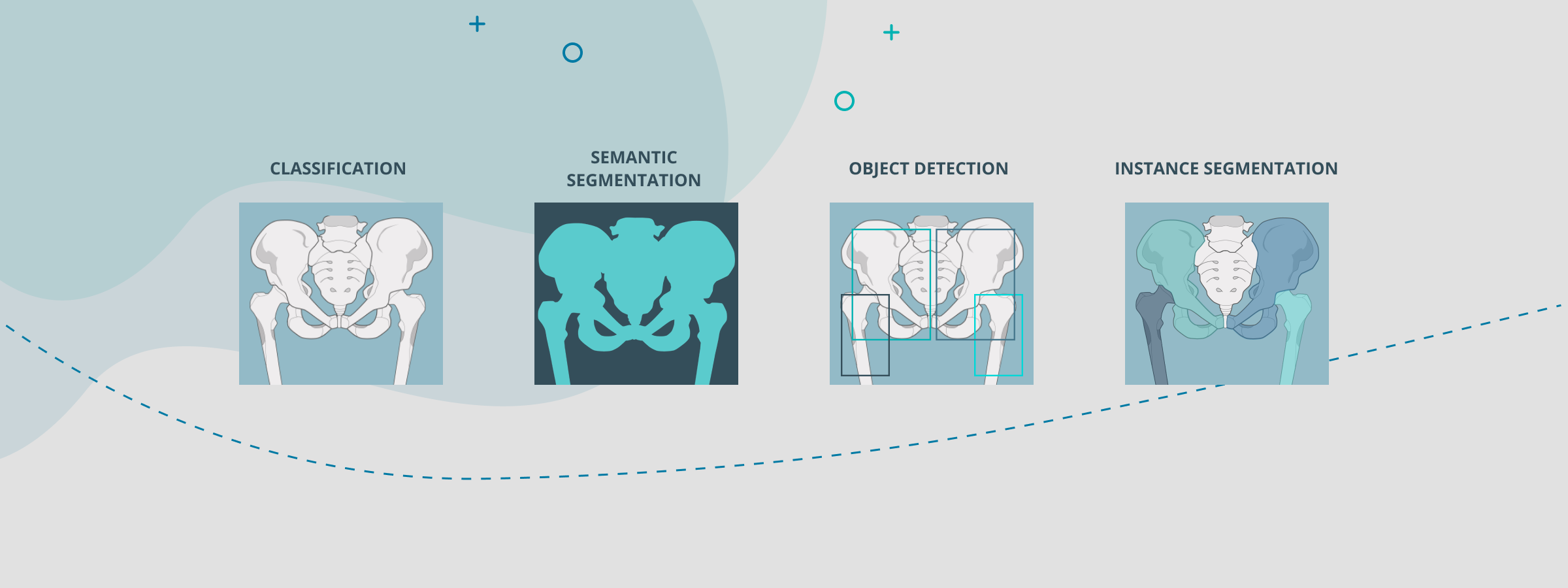
In general terms, image segmentation is a technique where a digital image is processed and analyzed into multiple parts or regions based on what object they represent.
Other techniques can often be used with segmentation, such as:
-
Image classification: In this task, the software attempts to identify the image into a category.
-
Object detection: It attempts to classify and locate objects in the input image. However, images are often selected and classified into a generic box.
There are different types of segmentation:
-
Semantic segmentation: Classifies each pixel of the image into a category. Semantic segmentation treats multiple objects within a single category or entity.
-
Instance segmentation: This is a much more detailed segmentation approach. It combines the advantages of segmentation and classification on a deeper level, by separating objects within the same category.
Another technique often used when you have a group of segmented images is image clustering in orthopedics. Essentially, it means that it becomes possible to cluster shoulder images and organize them into different groups. It would automatically place all the images with a long humerus to one side and a short humerus to the other.
Collections of objects are made through classification models, depending on their characteristics. Much as if they had different colors and the software would be organizing them.

How does image segmentation in orthopedics work?
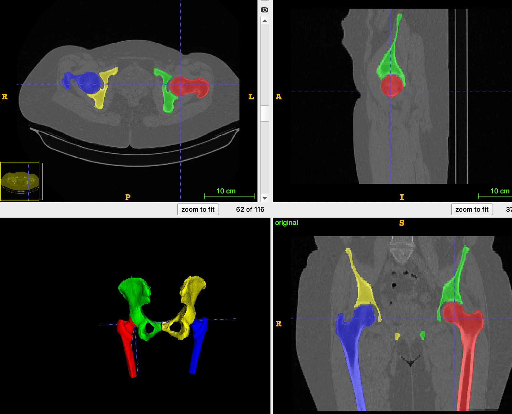
In orthopedics, image segmentation is a visual system that supports the surgeon when he is determining the strategy of how to assist a patient.
To make it simple, it works as a tool that allows surgeons to automatically select a specific bone and move it as they deem appropriate.
Image segmentation in orthopedics can play an essential role in medical image analysis. It allows the surgeon to better explore patient-specific anatomy without spending much time.
Take the example of the image below, where all the different regions/bones have been marked in different colors, allowing a better analysis of the patient-specific anatomy.
How does image segmentation work in orthopedics?
At PeekMed we've implemented top-notch technology that combines segmentation and Artificial Intelligence / Machine Learning.
If you haven’t read our article focused on AI in Healthcare and Orthopedics, let us remind you of the basics: Machine learning is a form of artificial intelligence. With it, computers learn from data without being programmed to complete a specific task.
What we do is use a process designated as instance segmentation. This makes it possible to classify subregions within a region of interest.
In practical terms, the technology can look at the x-ray and make sense of what is bone and what is not, thus facilitating the orthopedic surgeon process.
This is possible thanks to Deep Learning, as the algorithm takes the image and assigns importance to the various elements.
Let’s take a CT as a practical example of ct segmentation orthopedics.

After the images are captured in the CT scan machine, these are processed and converted into a DICOM file. Thus, becoming possible to combine it with artificial intelligence and, with it, sub-technologies such as computer vision.
Making it simple, the CT Image segmentation technique allows surgeons to select the bone almost automatically. You may think about image segmentation in orthopedics as the magic wand tool from Paint or Photoshop.
With a few clicks, the data points and object detection are automatically made and you may proceed with the clinical evaluation without much effort.
