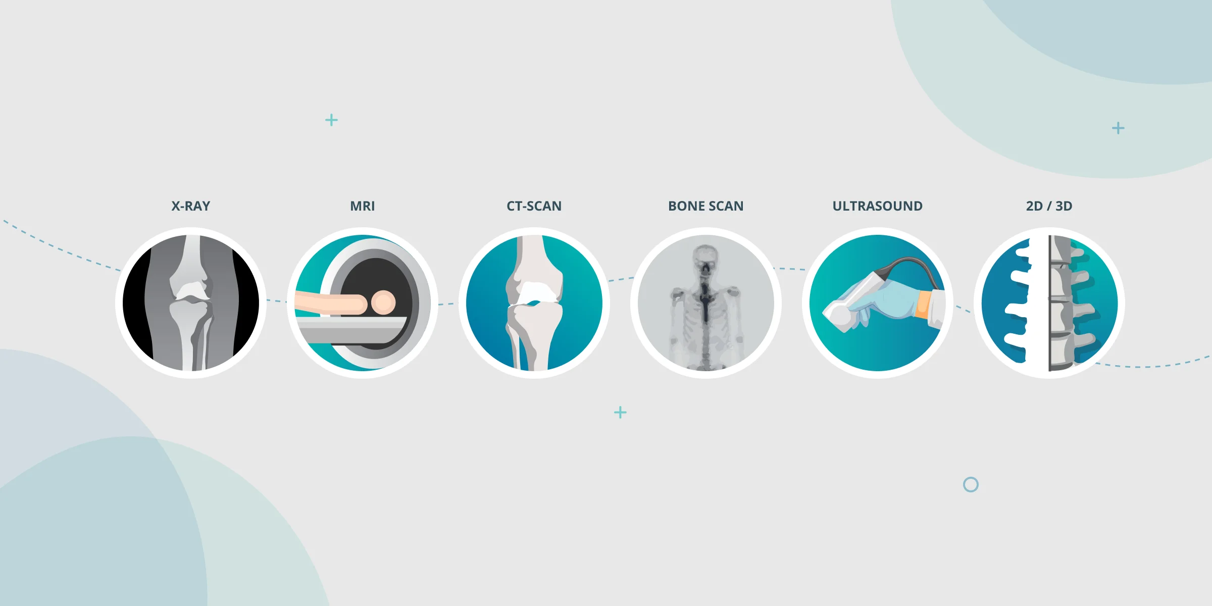
In orthopedics, accurate and detailed imaging plays a pivotal role in diagnosing, pre-operative planning, and monitoring various musculoskeletal conditions. With the advancement of technology, several imaging modalities have emerged, each with its advantages and disadvantages. Let’s explore the most common medical imaging techniques used in orthopedics.

X-ray Imaging
X-ray is the most widely used orthopedic imaging technique. It provides detailed visualization of the skeletal system by utilizing electromagnetic radiation.
Advantages:
- Quick radiographic images;
- Promptly assess fractures and identify their severity;
- Monitors fracture healing progress and evaluates the alignment of implants or joint replacements;
- Widely available and low cost, making it a cost-effective option;
- Simplicity and speed make it a convenient initial screening tool.
Disadvantages:
- 2D view, making it challenging to assess complex structures accurately;
- Soft tissues such as muscles, tendons, and ligaments are not visible;
- Bones can absorb radiation and block significant diagnostic data;
- Produces medium-quality images;
- Risk of excessive ionizing radiation exposure, especially when repeated frequently.

MRI Imaging
Magnetic Resonance Imaging (MRI) has revolutionized orthopedic imaging by producing detailed images of bones and soft tissues. It utilizes a strong magnetic field and radio waves to generate cross-sectional images of the body.
Advantages:
- Exceptional soft tissue visualization, making it ideal for assessing ligaments, tendons, cartilage, and other structures surrounding joints;
- Detection of soft tissue injuries, tumors, and infections, and evaluation conditions such as arthritis;
- Produces cross-sectional images with 3D capabilities;
- Non-ionizing radiation, making it a safer option for repetitive imaging;
Disadvantages:
- Expensive and high ongoing operation costs;
- Time-consuming;
- Less sensitive in detecting small amounts of calcification and bone fractures;
- Sensitive to patient movement, affecting image quality;
- Potential discomfort for claustrophobic and/or noise-sensitive patients;
- Interference with metallic implants or devices.
CT Scan Imaging
Computed Tomography (CT) scans employ a series of X-ray images taken from different angles to create detailed cross-sectional images of the body.
Advantages:
- Superior imaging of bone structures as it makes it clearer to identify bone overlap between images;
- Higher contrast resolution due to the expansion of the grey scale;
- Evaluation of complex fractures, bone tumors, and spinal conditions;
- Detailed 3D reconstructions, guiding surgical planning and orthopedic procedures;
- Faster than MRI;
Disadvantages:
- Exposure to ionizing radiation even higher than X-rays;
- Limited soft tissue contrast compared to MRI, making them less suitable for assessing soft tissue injuries or abnormalities;
- Sensitive to patient movement, affecting image quality;
- More expensive for the hospitals and the patients;

Bone Scan Imaging
Bone scintigraphy involves injecting small amounts of radiopharmaceutical with affinity to the bone into the bloodstream, which will follow a specific biodistribution throughout the whole body. Gamma cameras capture the radiation emitted, creating images highlighting areas of increased or decreased bone activity.
Advantages:
- High imaging sensitivity makes it easier to detect a wide variety of bone abnormalities such as tumors, fractures, infections, and bone metastasis.
- Whole-Body Scan or targeted evaluation of bone vascularization and regeneration (Three-Phase Bone Scan);
- Post-operative surveillance tool to access implant infection (aseptic loosening) or prosthesis detachment, indicating the potential need for revision surgery;
- Enhanced 3D bone imaging achieved through SPECT technique, often combined with CT (SPECT/CT), provides improved visualization of the region of interest.
Disadvantages:
- Time-consuming;
- Although painless, it is invasive;
- Sensitive to patient movement, affecting image quality;
- Low anatomical resolution;
- Although small, there is exposure to ionizing radiation;
- Lack of specificity, requiring extra imaging modalities for confirmation;
- Less availability in hospitals, compared to other imaging modalities.

Ultrasound Imaging
Ultrasound imaging uses high-frequency sound waves to produce real-time images of the musculoskeletal system. This allows the operator to discuss real-time scan findings.
Advantages:
- Cost-effective;
- Widely available and can be made portable;
- Non-ionizing radiation;
- Provides a clear image of soft tissues;
- Real-time assessment of joint movement and ligament and tendon injuries;
- Assists in minimally invasive procedures, like guiding injections or barbotage.
Disadvantages:
- Limited penetration which interferes with the visualization of deep structures, bones, or dense tissues;
- Operator dependence affects the quality of the image obtained;
- Not optimal for evaluating complex fractures or deep-seated pathologies.

3D Imaging Technology: Converting X-rays to 3D Bone Models
The quest for the best imaging technique continues in the ever-evolving field of orthopedics. While each modality has its pros and cons, an emerging solution combines traditional X-rays' strengths with the transformative power of advanced imaging technology: converting X-rays to 3D bone models.
Generating a 3D model introduces a new dimension of precision and planning that surpasses the limitations of traditional imaging in orthopedics. X-rays provide a cost-effective and widely available initial assessment, while 3D X-ray models allow for enhanced visualization of complex structures, overcoming the limitations of 2D X-rays and the high costs of MRIs and radiation exposure of CT Scans.
This visualization aids in identifying subtle abnormalities and optimizing implant placement. With this, orthopedic surgeons can enhance their diagnostic accuracy, streamline pre-operative planning, and improve patient outcomes.
Remember, when considering which imaging to order, it is crucial to evaluate the specific clinical scenario, the information required, and the risks associated.
