The Role of Advanced Imaging Technology in Orthopedics
You already know that imaging technology is essential in modern medicine. But did you know it's completely transforming orthopedics? As an orthopedic professional, you understand imaging technology's critical role in diagnosis, treatment planning, and surgical execution. However, with rapid advancements in technology, orthopedic imaging is more precise and efficient than ever before.
Imaging technology is crucial in modern orthopedics, enabling precise diagnosis, treatment planning, and surgical execution. Advances in imaging techniques such as X-ray, MRI, and CT scans have revolutionized orthopedic care, improving patient outcomes and streamlining clinical workflows.
Imagine having the ability to visualize complex fractures in three dimensions, accurately plan surgeries with AI-driven insights, and execute procedures with real-time imaging guidance. The best part? These innovations are already transforming orthopedic care.
In this guide, you'll discover how advanced imaging technologies—like X-ray, MRI, and CT scans—are revolutionizing orthopedic care. Plus, we’ll show you how PeekMed is leading the way in innovation.
Importance of Imaging in Orthopedics
Effective imaging is the foundation of accurate orthopedic diagnoses and successful treatment plans. It allows for precise visualization of musculoskeletal structures, ensuring better clinical decisions and improved patient outcomes. The integration of advanced imaging technologies ensures accurate assessments and enhances surgical precision.
Here’s a closer look at the key imaging modalities shaping modern orthopedics.
Key Imaging Modalities in Orthopedics
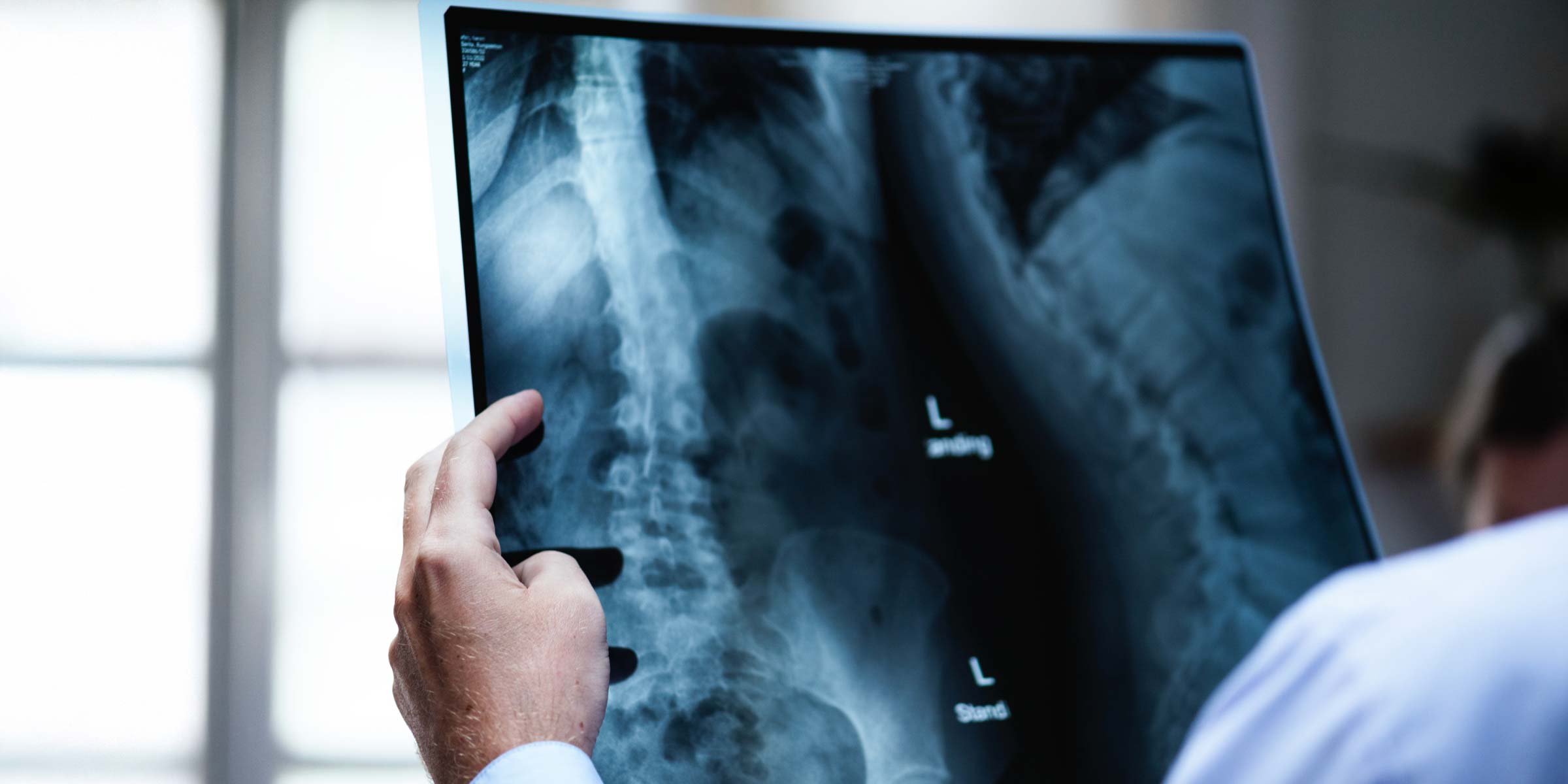
1. X-ray Imaging
X-ray remains the most widely used imaging modality in orthopedics. It is essential for detecting fractures, joint alignment, and bone deformities. The process of X-ray calibration ensures accuracy in image interpretation, minimizing errors in orthopedic diagnosis.
In other words, X-rays are the workhorse of orthopedic imaging. They quickly reveal fractures, joint alignment, and bone deformities.
But here’s the deal: X-ray calibration is crucial. Without it, even a small miscalculation can lead to misdiagnosis.
2. Magnetic Resonance Imaging (MRI)
MRI provides unparalleled soft tissue contrast, making it indispensable for assessing ligament tears, cartilage degeneration, and spinal pathologies. Its ability to capture high-resolution images without radiation exposure makes it particularly valuable for long-term monitoring.
Unlike X-rays, MRI does not use ionizing radiation, making it a preferred option for assessing chronic orthopedic conditions.
This means MRI is all about the details. It captures soft tissues—like ligaments and cartilage—with incredible clarity.
If you're dealing with a patient with a knee injury or spinal issue, MRI is your best friend.
3. Computed Tomography (CT) Scans
CT scans take imaging to the next level. CT scans offer high-resolution cross-sectional images of bones and joints, aiding in complex fracture assessment and preoperative planning. They create 3D cross-sections of bones and joints, making them a game-changer for complex fractures and surgical planning.
These scans play a pivotal role in 3D reconstructions. This is particularly beneficial in trauma cases, preoperative planning, and postoperative assessments.
Advanced Imaging Applications in Orthopedics
1. DICOM Imaging and Its Impact
DICOM (Digital Imaging and Communications in Medicine) enhances orthopedics workflows and enables seamless integration of imaging systems across different platforms, ensuring continuity in patient care. It is the industry standard for storing and sharing medical imaging data.
2. Image Segmentation for Precision Orthopedics
Advancements in image segmentation allow for detailed differentiation of anatomical structures, improving AI-assisted diagnostics and personalized treatment plans. This technology enhances preoperative planning by isolating bones, soft tissues, and implants with high precision.
3. Orthopedic-Specific Imaging Technology
Specialized imaging techniques, such as weight-bearing CT scans and fluoroscopy, provide deeper insights into joint function and biomechanics. These technologies are crucial for evaluating orthopedic conditions that traditional imaging methods may overlook.
The Role of Imaging in Surgical Planning and Execution
Preoperative Planning
Advanced imaging provides orthopedic surgeons with critical preoperative insights, reducing uncertainties before entering the operating room. 3D CT reconstructions and AI-driven simulations allow for precise surgical mapping, minimizing risks and improving outcomes.
Intraoperative Imaging
Real-time imaging techniques, such as fluoroscopy and intraoperative CT, act as navigation tools during surgery. These imaging modalities enhance precision in implant placement, fracture fixation, and minimally invasive procedures, leading to reduced operative time and improved patient safety.
Research shows that integrating advanced imaging into surgical workflows enhances accuracy and decreases procedural complications.
Postoperative Assessment
After surgery, imaging plays a crucial role in monitoring healing, assessing implant positioning, and detecting complications. Emerging technologies like AI-assisted imaging analysis and augmented reality are further refining postoperative evaluations, improving long-term patient outcomes.
PeekMed: Revolutionizing Orthopedic Imaging and Surgical Planning
PeekMed integrates cutting-edge imaging technology to streamline orthopedic planning and execution. By leveraging AI-powered 3D modeling and advanced imaging analytics, PeekMed enhances preoperative simulations, reducing surgical risks and optimizing patient outcomes.
Key Benefits of PeekMed:
- 3D Surgical Planning: Converts 2D scans into 3D models for precise preoperative visualization.
- AI-Assisted Image Analysis: Enhances diagnostic accuracy and treatment recommendations.
- Seamless DICOM Integration: Ensures compatibility with various imaging platforms.
Advanced imaging technologies are indispensable in orthopedic diagnostics, treatment planning, and surgical execution. By integrating AI-driven imaging solutions like PeekMed, orthopedic professionals can enhance patient care, improve surgical precision, and patient outcomes, and optimize clinical workflows.
As imaging evolves, staying ahead of these advancements will be key to delivering better, faster, and safer orthopedic care.
References
- Sharma, G., Parvizi, J., & Kwon, Y. M. (2021). Artificial intelligence in orthopedic surgery: Current state and future perspective. Journal of Orthopedic Research, 39(7), 1363-1375. PubMed
- Jentzsch, T., Vlachopoulos, L., & Fürnstahl, P. (2023). Advanced imaging in orthopedic surgery: Applications and future directions. Clinical Orthopedics and Related Research, 481(1), 15-29. ScienceDirect
- Taylor, S. A., & Amanatullah, D. F. (2022). Intraoperative imaging in orthopedic surgery: Benefits, limitations, and new developments. Journal of Bone and Joint Surgery, 104(5), 432-444. PMC
- Sanders, J. O., & Little, D. G. (2021). The impact of 3D imaging on orthopedic practice: A systematic review. Orthopedic Clinics of North America, 52(4), 581-596. PMC
