PeekMed
If you’ve ever stepped into an operating room, you know the truth: you can’t treat what you can’t see.
As surgeons, precision is everything. We need more than a diagnosis. We need a clear, accurate map of the patient’s anatomy.
The good news? Medical imaging technology gives us that map. In stunning detail.
In this article, we’ll break down what medical imaging technology is, how it works, and why it’s reshaping modern surgery, especially in orthopedics.
Defining Medical Imaging Technology
Medical imaging technology refers to the methods and tools that let us see inside the body without surgery.
Think of it as the surgeon’s “X-ray vision”, powered by advanced physics, digital processing, and often, artificial intelligence.
From X-ray calibration to 3D reconstructions, these tools help bridge the gap between diagnosis and surgical precision.
From X-rays to AI-assisted Planning
The first X-ray was taken in 1895. It was a game-changer.
Today, we have:
- CT scans: Multiple images combined into a 3D model.
- MRI: Detailed views of soft tissue without radiation.
- Ultrasound: Real-time imaging for moving structures.
- Specialized orthopedic imaging for precise bone and joint mapping.
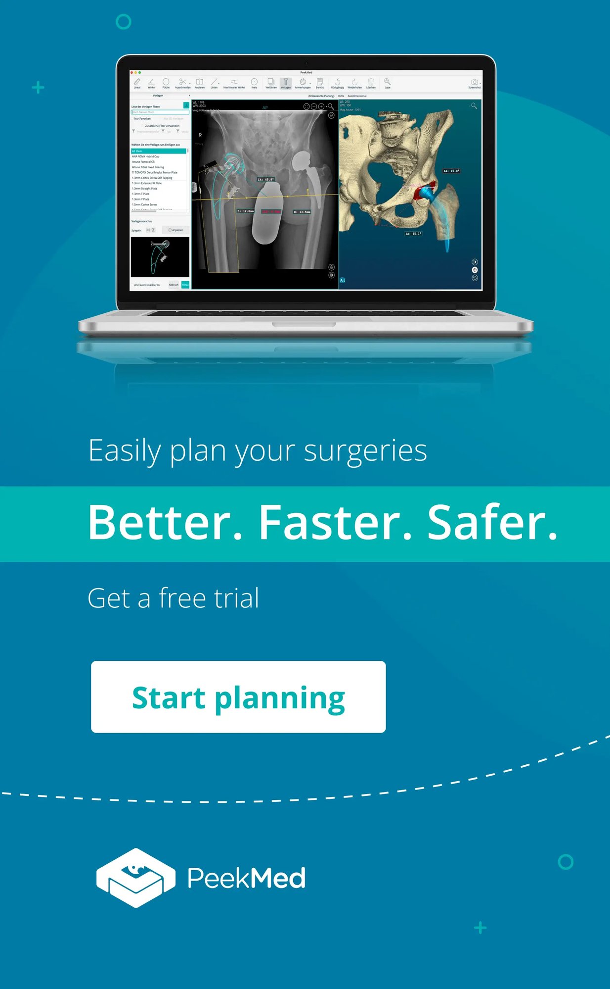
The real leap forward? Integrating these scans into platforms like PeekMed so surgeons can simulate and plan procedures before entering the operating room.
How Medical Imaging Works
No matter the type, all imaging shares one principle: energy interacts with tissues, and sensors capture that interaction.
- X-ray: Radiation passes through the body, highlighting dense structures like bone.
- MRI: Magnetic fields and radio waves reveal soft tissues.
- CT scan: Multiple X-rays from different angles create detailed cross-sections.
- Image segmentation isolates specific structures for precise study.
Modern systems don’t just capture static images; they generate dynamic, interactive 3D models we can measure, rotate, and plan around.
Why It Matters in Surgery
Picture a patient with a complex fracture. Without imaging, every decision in surgery is a guess.
With imaging, we can:
- Trace fracture lines exactly
- Measure distances down to fractions of a millimeter
- Plan implant placement virtually before the first incision
Integrating DICOM images into surgical planning helps anticipate challenges, shorten procedures, and improve outcomes.
Orthopedics: Precision by Design
In orthopedic surgery, accuracy isn’t optional. It’s essential. Whether reconstructing a joint or correcting a deformity, precise imaging is our guide.
That’s why imaging technology in orthopedics is advancing so quickly and why platforms like PeekMed are becoming a standard tool in preoperative workflows.
The next chapter of medical imaging technology is about more than seeing.
It’s about predicting outcomes, simulating procedures, and guiding interventions in real time. AI, automation, and augmented reality are already changing the way we operate, and the trend is only accelerating.
Medical imaging technology is the eyes of modern medicine. When combined with surgical planning platforms like PeekMed, it becomes the surgeon’s most powerful ally for delivering safe, precise, and efficient care.
Explore More on Medical Imaging
If you found this overview helpful, you might want to dive deeper into specific areas of medical imaging technology:
- 📌 DICOM Images – Understand the standard file format behind medical imaging and why it’s essential for interoperability.
- 📌 X-ray Calibration – Learn how precise calibration ensures accurate diagnostics.
- 📌 Image Segmentation in Orthopedics – See how AI and algorithms separate structures for clearer surgical planning.
- 📌 Orthopedic Imaging – Discover imaging methods designed specifically for bones and joints.
- 📌 Imaging Technology in Orthopedics – Explore the innovations shaping orthopedic surgery today.
These resources connect directly to our Medical Imaging Technology Pillar Page, your go-to guide for understanding how imaging is revolutionizing patient care.


